Micro-CT for biology, zoology, and archaeology
Morphological examination via micro-CT: bones, soft tissue, and fossils
You want to examine the interior of your biological samples or archaeological finds without damaging them. As an accredited CT scanning service provider from Germany, we perform CT examinations in industrial environments as well as CT scans on biological samples as an alternative to histology. Using 3D X-ray examination, we deliver up to 1,000 tissue sections per millimeter without physically touching your precious samples. We also provide microscopic 3D data (micro-CT and nano CT) for archaeological finds with a resolution of up to 1 µm – your samples remain optimally protected, as the CT examination is contactless and non-invasive.
Your advantages with the micro-CT & nano CT services from TPW CTinspect:
- Your TPW expert: Dr. Thomas Kleinteich – Doctor of Zoology (CT muscle fibers, bone FEA) as your sparring partner for parameters, preparation, and measurement
- Non-destructive: preservation of rare samples, ideal for museums and collections.
- Microscopic resolution as required: Micro-CT up to Nano CT (max. 1 µm)
- Analyzing soft tissue: Advice on contrasting organs and muscle fibers
- Fast results: Less effort than histology. Permanent digital volume data for virtual 3D histology
- Data sovereignty & compliance: Data storage in Germany, password-protected download area
- On-site support in Neuss, Germany: Live participation on scan day possible and joint data analysis with our CT engineers
What you get from us: Data, visualizations, reports
CT volume data (3D data)
Digital image data
Data also available in STL format on request (e.g., for 3D printing/3D reconstructions)
Report with cross-sectional images and evaluation (upon request):
Segmentation (e.g., CT bone/soft tissue)
Fiber orientation (false colors)
Metrics (porosity, dimensions, trabecular analysis)
TPW CTinspect also provides assistance with preparation and contrast enhancement
Your CT expert for biological samples: Dr. Thomas Kleinteich

- Sparring partner for industrial computed tomography scanning, including in the biological/zoological field, thanks to years of practical experience
- Doctorate in zoology, including CT analysis of muscle fibers in vertebrates
- FEA for the simulation of bone anatomy based on computed tomography data
- Publications in the field of CT analysis of biological samples (selection):
- E. W. M. Paig‐Tran, T. Kleinteich, and A. P. Summers, “The filter pads and filtration mechanisms of the devil rays: variation at macro and microscopic scales,” Journal of Morphology, vol. 274, no. 9, pp. 1026–1043, Sep. 2013.
- A. van der Meijden, T. Kleinteich, and P. Coelho, “Packing a pinch: functional implications of chela shapes in scorpions using finite element analysis,” Journal of Anatomy, vol. 220, no. 5, pp. 423–434, May 2012.
- T. Kleinteich, H. C. Maddin, J. Herzen, F. Beckmann, and A. P. Summers, “Is solid always best? Cranial performance in solid and fenestrated caecilian skulls,” Journal of Experimental Biology, vol. 215, no. 5, pp. 833–844, Mar. 2012.
- T. Kleinteich, A. Haas, and A. P. Summers, “Caecilian jaw-closing mechanics: integrating two muscle systems,” Journal of the Royal Society Interface, vol. 5, no. 29, pp. 1491–1504, Dec. 2008.
- T. Weihmann, T. Kleinteich, S. N. Gorb, and B. Wipfler, “Functional morphology of the mandibular apparatus in the cockroach Periplaneta americana (Blattodea: Blattidae) - a model species for omnivore insects,” Arthropod Systematics & Phylogeny, vol. 73, pp. 477–488, Dec. 2015.
- T. Kleinteich and S. N. Gorb, “Frog tongue surface microstructures: functional and evolutionary patterns,” Beilstein Journal of Nanotechnology, vol. 7, no. 1, pp. 893–903, Jun. 2016.
- T. Kleinteich, F. Beckmann, J. Herzen, A. P. Summers, and A. Haas, “Applying X-ray tomography in the field of vertebrate biology: form, function, and evolution of the skull of caecilians (Lissamphibia: Gymnophiona),” in Developments in X-Ray Tomography VI, vol. 7078, pp. 113–122, Sep. 2008.
- K. Engelkes, J. Helfsgott, J. U. Hammel, S. Büsse, T. Kleinteich, A. Beerlink, S. N. Gorb, and A. Haas, “Measurement error in μCT-based three-dimensional geometric morphometrics introduced by surface generation and landmark data acquisition,” Journal of Anatomy, vol. 235, no. 2, pp. 357–378, Aug. 2019.
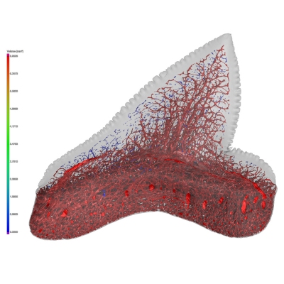
CT of muscle fibers in zoological samples – Micro-CT & Nano CT for morphology
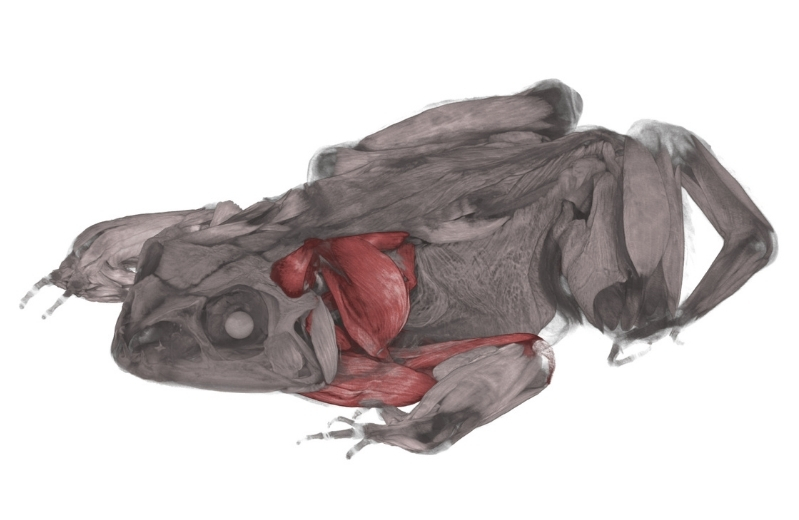
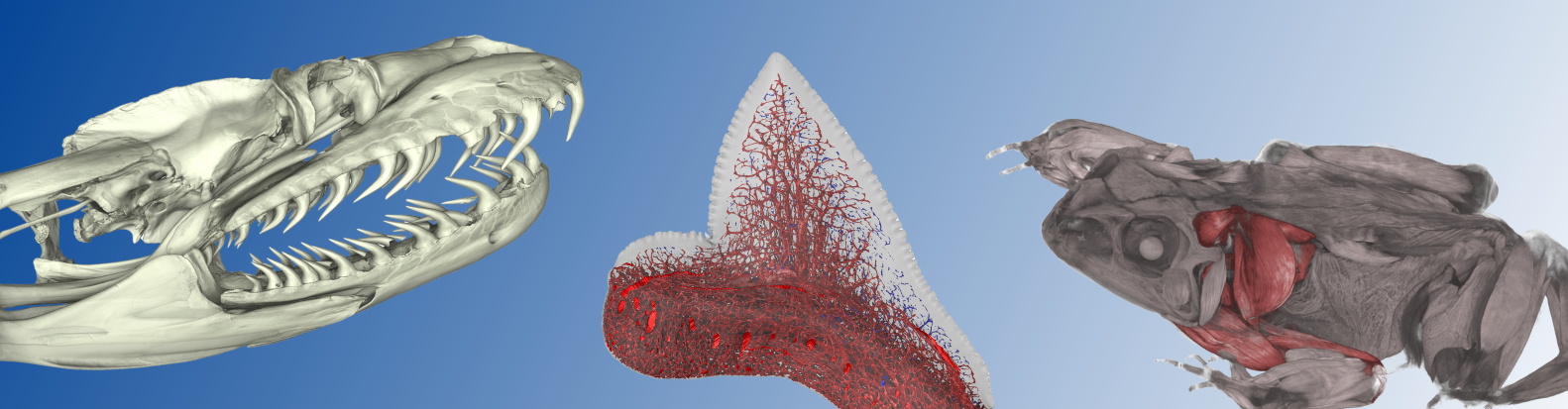
Biological soft tissues, such as muscle fibers, are often very complex and can only be understood in a three-dimensional context with other anatomical structures. With micro CT and nano CT, we can visualize and examine this three-dimensionality in a non-destructive manner. Soft tissues can be made visible in a targeted manner using suitable contrast agents, and we are happy to advise you on this. This allows the fiber orientation and fiber architecture within the specimen to be traced.
Computed tomography of zoological muscle fibers thus provides insights that a classic series of thin sections can only deliver with considerable effort. As an alternative to histological examination, this creates a virtual 3D histology that leaves the specimen intact. Select the number of sections and section planes as often as you like, purely digitally. This is also particularly valuable when samples serve as the basis for finite element analysis (FEA), for example in functional questions.
We are happy to advise you on preparation (dry/in liquid) and fixation. During a joint examination on site in Neuss,Germany, we can precisely define your area of interest and the artifacts to be examined. We are aware of the rarity of biological samples, which is why we coordinate parameters and the handling of your samples closely with you. Benefit from our expertise in zoology (including CT scanning of muscle fibers and bones). Dr. Kleinteich is your sparring partner for optimal imaging.
CT scan archaeology: fossils and bones
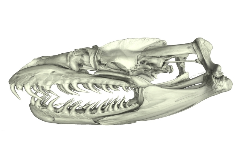
The examination of fossils, bones, and archaeological objects is greatly simplified by the imaging technique of micro-CT. Our 3D X-ray archaeology provides a non-destructive view of the interior and reveals structures such as anatomy, bone fractures, trabeculae, inclusions, and manufacturing techniques. Structures can also be made visible in sealed objects or in a matrix of clay or earth. This is less risky than mechanical exposure. Our CT scanning service provides volume data for virtual measurements and 3D reconstructions (e.g., surfaces as STL data for 3D printing). For museums and research institutes, this means that sensitive objects remain intact and samples can continue to be used. The 3D data obtained forms a solid foundation for publications, knowledge transfer, and restoration.
Insects & invertebrates in micro-CT – morphology in 3D
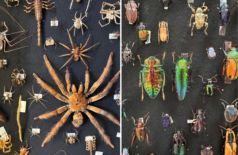
The fine structures of insects and other invertebrates can be excellently visualized using micro-CT. This makes the structures of the head, mesothorax, and metathorax visible in three dimensions. Virtual preparation replaces risky mechanical interventions and time-consuming preparation in casting resin, as layers, segments, and cavities can be separated digitally and in all spatial directions. If required, we are happy to advise you on the preparation of samples to emphasize soft tissue. For very small objects, nano CT is used to visualize structures down to a size of 1 µm. For natural history museums and research teams, this means precise results and continued pristine examination objects.
Our data sets are suitable for geometric morphometry, taxonomy, and the creation of vivid 3D models.
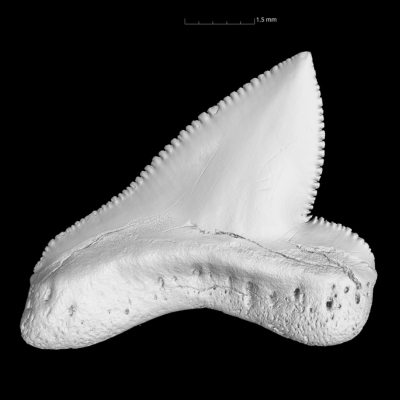
Teeth & dental structures using micro CT scanning
Micro CT scanning is ideal for imaging teeth and dental structures. It can be used to detect cracks, root canals, and abrasions. The volume data obtained enables precise measurements, virtual preparations, and 3D reconstructions, which are suitable for research and knowledge transfer.
Selection of possible sample types:
We can perform three-dimensional industrial CT scanning of a wide range of zoological samples or archaeological finds using computer tomography – here is a selection:
• CT muscle fibers
• CT soft tissue/organs
• CT bones
• CT analysis of trabecular structures
• CT skeletal structures
• CT fossils
• CT anatomy
• CT corals
• CT teeth
• CT insects and invertebrates
About TPW CTinspect:
About us
Learn more about the industrial CT scanning experts of TPW CTinspect from Neuss, Germany..
LEARN MORE
Accredited testing laboratory
More about our certificates as an accredited testing laboratory for CT services...
LEARN MORE
Your contact
Our TPW expert Dr. Thomas Kleinteich in the field of CT scanning of biological samples and CT archaeology...
LEARN MORE